

Detailed Imaging with Musculoskeletal Ultrasound
Discover the Source of Your Pain Quickly and Reliably!
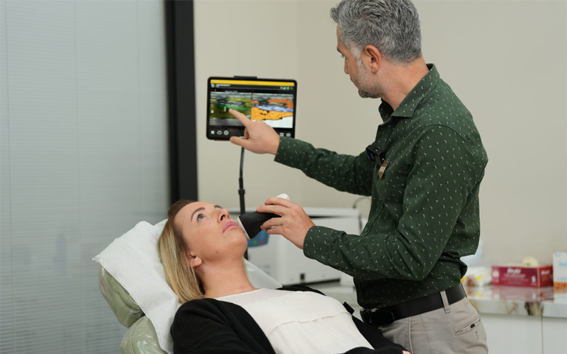
Musculoskeletal ultrasound is a non-radiative, safe diagnostic method that allows detailed imaging of muscles, tendons, ligaments, joints, and soft tissues. With real-time imaging, it provides the ability to examine structures in motion and helps ensure accurate diagnosis and effective treatment planning.
What is Musculoskeletal Ultrasound?
Musculoskeletal ultrasound is a safe, non-invasive technology that uses high-frequency sound waves to visualize the body's internal structures. It is particularly useful for evaluating the following:
-
Muscles
-
Tendons
-
Ligaments
-
Joint capsules
-
Bursae
-
Nerves
When is it Used?
-
Muscle and Tendon Injuries: Muscle tears, Tendon ruptures, Tendinitis (inflammatory conditions)
-
Ligament and Joint Issues: Sprains, Ligament damage, Joint effusion (fluid accumulation in joints)
-
Nerve Compression: Carpal tunnel syndrome, Other peripheral nerve compressions
-
Cysts and Masses: Ganglion cysts, Lipomas, Other soft tissue masses
-
Injection Guidance: Ensures accurate and precise placement of therapeutic injections.
Advantages of Musculoskeletal Ultrasound
-
Real-Time Imaging Dynamic structures can be observed, offering functional assessment opportunities.
-
Radiation-Free and Safe Because it does not use X-rays or magnetic fields, it is safe for all age groups.
-
Fast and Effective Instant results provide accurate diagnoses with no waiting time.
-
High Resolution Provides clear, detailed images of soft tissues for precise evaluation.
-
Suitable for Pregnant Women and Implant Patients As it does not involve radiation, it is an ideal imaging method for pregnant women and patients with metal implants.